|
|
|
Program > • Session New instrumental developments and techniquesSince the very first meeting in Como in 1999 many developments in instruments and fluorescent dyes have moved from looking from colonies down to single cell and in the cell (genomics, proteomics). The analyses are made at a frequency out of reach of most of the techniques available until then. These high throughput screening of cultures or samples allow at the same time the isolation of rare populations thanks to optical resolution of the cells of interest and physical isolation by cell sorting. New light sources are available to extend the range of excitation of the particles (cells). More and more colors can be simultaneously collected and discriminated. Some instruments are also able to collect the whole signal (light scatter and fluorescence intensities) along the particles, providing unique fingerprints of the cells. With the new generations of instruments, up to six populations can be sorted in the same run. With always improved technical performance, constant decrease in cost of the instruments, continuous increase in computing power and data storage, classical image cytometry is on its way, providing a unique opportunity to convert dots on a cytogram into a real picture of the cell. The last developments have come along in the in situ analysis and imaging of aquatic microbes (phytoplankton) with the Flow Cam or the Cytobuoy instruments. Unlike the concept of static imaging or scanning systems these instruments really deserve the name of "image flow cytometer" as cells are imaged as they flow in the instruments. Whilst the small flow channels in most instruments are already justify the name micro fluidic device there are still developments under way for small and disposable micro fluidic instruments. Chairman : Prof. J. Paul Robinson (Purdue University Cytometry Laboratories, Lafayette, USA) 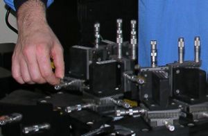
|
| Online user: 1 |

|
